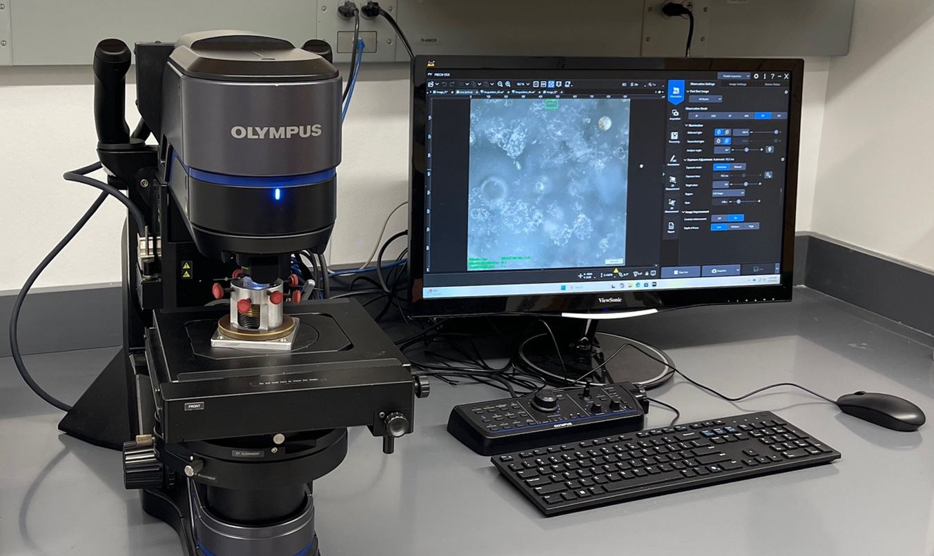
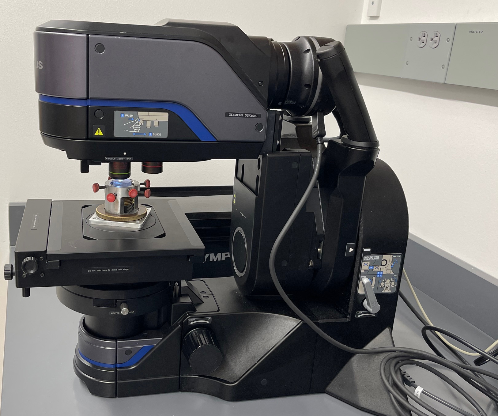
Available Methods and Accessories
- 1x super long distance objective for stereoscope imaging
- 5x Objective (up to 10x zoom, analogous to analog 50x objective)
- 20x Objective (up to 10x zoom, analogous to analog 200x objective)
- Bright field, Oblique, Dark field, Mixed Bright/Dark field, Polarized, and DIC lighting
- Motorized Z-head
- Motorized, rotatable sample stage
- Tilt head for oblique imaging
Sample Requirements
- Up to 4” height
- 6” x 6” length x width
Summary of Technique
Microscopy centers around visualization of regions of interest on the micrometer scale. Bright field microscopy is a well known type of viewing for optical microscopy, allowing the detection of reflected light from a sample surface to show defects or features that impact materials. Oblique lighting shines light onto a sample from an angle, allowing for shadows to be cast and detect surface features that would otherwise be obscured by direct lighting conditions. Dark field microscopy shines light on the edges of the sample, allowing the scattered light to reflect from scratches and cracks more easily. Polarized microscopy utilizes two filters, a polarizer and an analyzer, to detect changes in the rotation of light which can be related to material properties (crystal structures, birefringence, etc). Differential interference contrast lighting shears the reflected polarized light so that samples normally difficult to image under bright field applications are more easily viewed in high contrast.
Information Provided & Detection Limits
Micrographs provide detailed images of microscopic features on samples. Angle information from the stage, tilt head, and polarizers is available for data logging and analysis.
Lab Location and Contact Information
Lab Location: Science 1, G19 (Mechanical Testing Lab)
Lab Manager: Nicholas Eddy
nicholas.eddy@uconn.edu
860-486-2568

